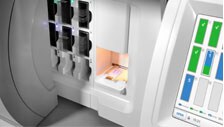
Digital pathology, which can also be referred to as virtual microscopy, involves the capturing, managing, analyzing and interpreting of digital information from a glass slide.
The process involves glass slides and converting these to digital pathology slides using digital pathology scanning solutions. A digital slide image is then generated which allows for high resolution viewing, interpretation and image analysis of digital pathology slides.
By leveraging the value of digital pathology imaging, we are delivering on a vision of precision medicine.
Where do we fit in?
By transforming the digital pathology landscape, we’re able to accelerate progress on the research that matters impacting billions of people globally. As market leaders in digital pathology workflow software serving over 40,000 users worldwide, our reach spans many sectors and application areas.
Things to consider when investing in WSI scanners for pathology
Improving pathology
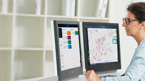
Our deep expertise in web-enabled workflow, case management, research collaboration and cellular image analysis software convinces us that these tools can enhance pathology in a variety of critical areas.
The right tools for pathologists
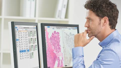
In its present form, healthcare is largely reactionary. Personalized medicine, tailoring treatment of a patient based on his or her unique physiological characteristics, will change the practice of medicine. It will allow healthcare providers to anticipate on and prevent disease progression by considering genetic and environmental factors that may increase predisposition.
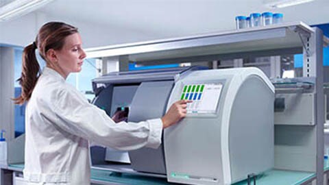
Pathology & the future of personalized medicine
The full scope of personalized medicine has not yet come to fruition, but it is already influencing cancer care.
Appropriate therapeutic regimens for various cancers are being determined by molecular testing. Tumors might present as a textbook example of a certain type but with molecular techniques like florescence in situ hybridization (FISH) technology, might actually be revealed as a completely different type of lesion.
Philips Digital & Computational Pathology is committed to play its role in helping pathologists reach the ultimate goal of changing healthcare by allowing them to see more and share knowledge.
