
FieldStrength MRI magazine
News - June 2021
MRI becoming a realistic option in prenatal diagnosis of cardiac disease
The possibility to obtain a reliable fetal cardiac gating signal presents the exiting opportunity to perform robust fetal cardiac MR as an additional means for fetal congenital heart disease diagnosis. For example, when fetal cardiac echocardiography is inconclusive, MRI studies of heart anatomy and function can provide valuable information to aid in prenatal diagnosis and in preparing for surgery after delivery. Previously, fetal cardiac gating was not available, but the recently developed Doppler ultrasound MR gating device now facilitates gated fetal cardiac MR studies. The gating device is compatible with Philips MRI scanners and connects wirelessly, without need for special software installation. It may also be used for adult and pediatric cardiac triggered scans.
“The direct gating with ultrasound will be a game changer for the clinical use of fetal cardiac MRI”
Mike Seed,
The hospital for Sick Children, Toronto
Fetal cardiac MR imaging with smart-sync
Typically, cardiac MR utilizes either electrocardiography (ECG) or vectorcardiography (VCG) techniques to synchronize MR acquisition with the heart rhythm to “freeze” heart motion. Both methods require the attachment of electrodes to the patient’s skin, which makes the approach unsuitable for fetal cardiac imaging. The recently introduced Doppler ultrasound device for gating, smart-sync, was developed by northh medical GmbH in Germany, in collaboration with Philips and clinical collaborators. This device can detect the heartbeat during MRI acquisition, and sends a gating signal to the scanner. This allows for the synchronization of MR data acquisition with the fetal heartbeat, resulting in fetal cardiac MR images of diagnostic quality. It allows for the use of standard cardiac sequences, so that acquired images instantly appear on the screen, allowing for immediate evaluation of slice positioning and quality – an important feature that allows for adjustments should the fetus change position. High image quality can be obtained for morphological images as well as 2D and 4D blood flow measurements, in a normal clinical setting. Together with the acquisition speed of the Philips Compressed SENSE method, fast and well-directed imaging of the fetal heart and flow can be performed, opening up a new avenue for congenital heart disease diagnosis before birth. The ultrasound-based trigger signal is reliable and easy to use due to its wireless connection. The signal also remains undistorted by the MR field, unlike ECG and VCG methods, and eliminates the need to attach electrodes to the skin. The positioning of the smart-sync probe is quite simple, only requiring the proper placement above the fetal heart.
The exciting alternative option of using Doppler ultrasound for MR cardiac gating allows to synchronize MR image acquisition with the fetal cardiac rhythm in a way that was previously not possible. Without this synchronization, motion artifacts typically render cardiac MR images useless for diagnosis.
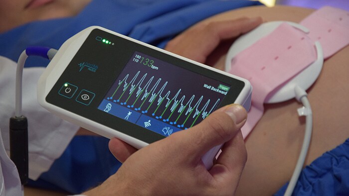
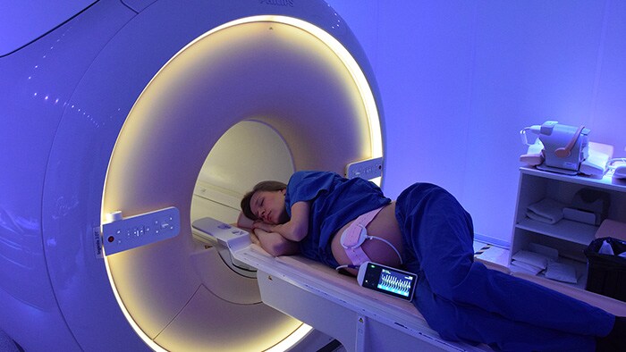
The transducer is fixed with a belt at the position where the fetal heart signal is strongest. The signal of the beating fetal heart is shown on the display and transferred wirelessly to the user interface.
“The fetal cardiac gating allows us to obtain MR images of the fetal heart with good diagnostic quality.”
Dr. Vanessa Berger-Kulemann, Radiology specialist
A useful alternative for prenatal echocardiography
However, since accurate prenatal diagnosis is closely associated with optimal postnatal outcomes, a first-time right diagnosis is highly desirable, especially in identifying cases of congenital heart defects. Therefore, the use of cardiac MR with smart-sync represents a long-awaited alternative for fetal ECG.
Typically, prenatal cardiac examinations are performed with ultrasound echocardiography. However, this technique can be limited in cases of late gestational age, due to calcification of the bone, fetal position, or a lack of amniotic fluid. In addition, echocardiography is highly observer-dependent and with no alternative methods for assessing inconclusive results.
“We have incorporated fetal cardiac gating into our clinical fetal CMR program, helping us in diagnosis of cardiac malformation in cases where fetal echocardiography was inconclusive.”
Pediatric Radiologist in a European university hospital
Fetal cardiac imaging with standard cardiac MR sequences
These sequences allow for immediate assessment of acquired images:
Cardiac MR in adults and children without need for electrodes
In essence, smart-sync solves these issues by measuring actual blood flow and cardiac motion via ultrasound. This allows for the acquisition of high quality cardiac images, as well as use of high temporal and spatial resolution, with notable benefits for quantification and longitudinal studies. Trigger signals based on ultrasound can diminish the number of incorrect signals, and circumvent the need for repeating scans due to motion artifacts.
Smart-sync gating can also be used in cardiac MR examinations for adults and children. It offers an undisturbed trigger signal, even at high fields, and eliminates the need for the unwieldly positioning of the adhesive electrodes on the patient’s chest. Conversely, conventional ECG/VCG is patient-dependent, is prone to electric field errors, and susceptible to arrhythmia, which particularly affects image acquisition in diastole, where the required delay can only be predicted and not measured, resulting in inaccurate predictions of arrhythmic heart beats.
“Smart-sync provides the cardiac gating signal that is necessary to run standard cardiovascular MR sequences with diagnostic quality.”
PD Dr. Björn Schönnagel, Radiology specialist
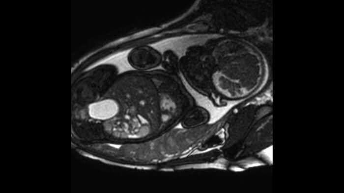
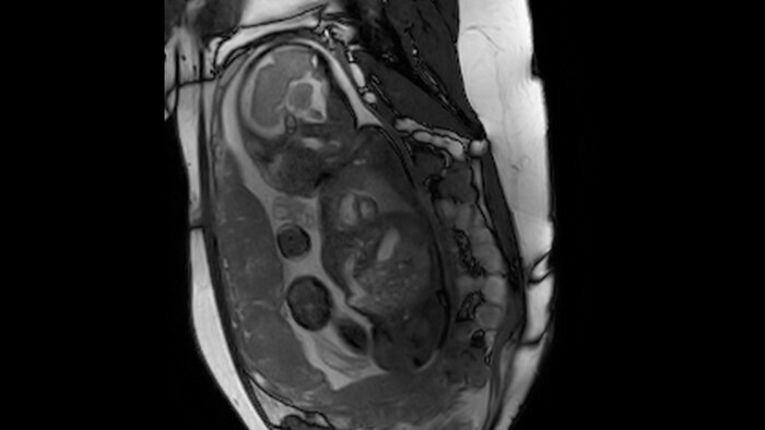
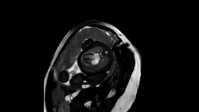
Imaging examples
Fetal cardiac imaging examples, gestational age 33-36 weeks.
Short axis view, 1.5T
Short axis view, 3.0T
Four chamber view, 1.5T
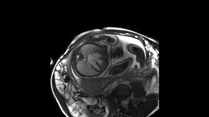
Four chamber view, 3.0T

4D Flow, 1.5T
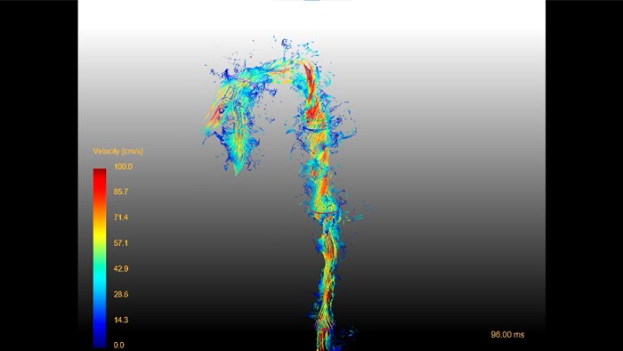
4D Flow, 3.0T
“The application of smart-sync for fetal cardiac MRI allows the translation of standard cardiac examination to the prenatal examination.”
Prof. Dr. Jin Yamamura, Radiology specialist
Wirelessly compatible with Philips MRI systems
Smart-sync can wirelessly connect to Philips 1.5T and 3.0T MRI systems, without the need for additional software. It has the potential to facilitate wide-scale availability of fetal cardiac MR.
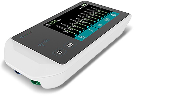
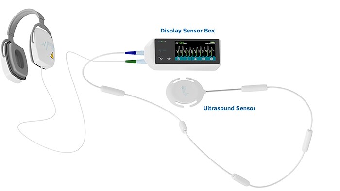
The smart-sync Doppler ultrasound components
The ultrasound sensor is placed above the fetal heart. The display sensor box shows the heartbeat pattern and generates a trigger signal, which is sent wirelessly to the Philips MRI scanner. The display incorporates a touch screen to switch between the fetal and adult application and to change settings. Headphones provide acoustic feedback to help with the proper positioning of the ultrasound sensor.
ExamCards for fetal cardiovascular MR with ultrasound gating device
These 1.5T and 3.0T ExamCards include basic MR sequences for studying the morphology and function of a fetal heart, based on cardiac gating via the smart-sync Doppler ultrasound device. The ExamCards may be used to help users familiarize themselves with the technique, and can be easily integrated into clinical procedures.
")
")
The sequences are balanced for both spatial (1.0 to 1.4 mm) and temporal (approx. 15 ms) resolution to accommodate fetal cardiac anatomy and physiology.
Compressed SENSE is included for scan time reduction.
For free-breathing scans, it is recommended to instruct shallow and regular maternal breathing. In addition, MultiTransmit with RF volume shimming at the fetal heart is advised on 3.0T for a tailored result of signal homogeneity.
These ExamCards are developed in collaboration with University Medical Center Hamburg-Eppendorf, Germany and Allgemeines Krankenhaus der Stadt Wien, Austria.
References
Results from case studies are not predictive of results in other cases. Results in other case may vary.

Subscribe to FieldStrength
Our periodic FieldStrength MRI newsletter provides you articles on latest trends and insights, MRI best practices, clinical cases, application tips and more. Subscribe now to receive our free FieldStrength MRI newsletter via e-mail.
Stay in touch with Philips MRI