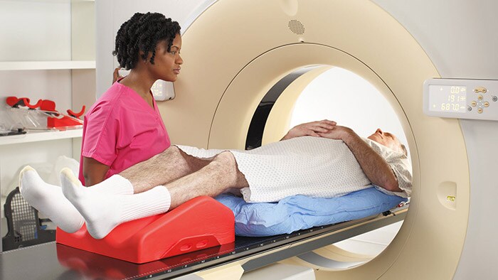
The post-processing of oncology image datasets (CT, PET/CT, and MR) can pose a major challenge for radiology and oncology staff. Numerous manual actions are often required to handle the significant amount of data produced by multiple follow-up studies. The Multi Modality Tumor Tracking application (MMTT) on IntelliSpace Portal provides the tools to simplify the review and analysis of CT, PET/CT, SPECT/CT and MR datasets for tumor detection and monitoring. This material is not for distribution in the USA
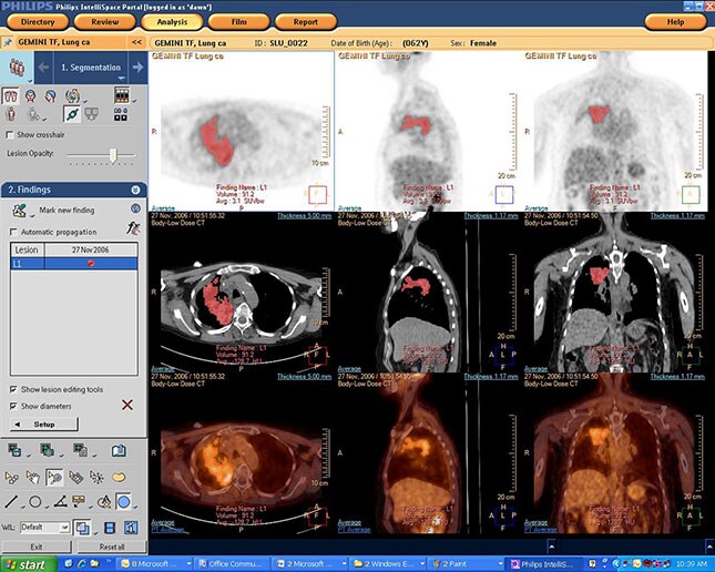
MMTT supports datasets from CT, MR, and PET/CT. The above image is of a 62-year-old female with a history of lung cancer. A large lesion is segmented in the right upper lobe. Lesion statistics were obtained for PET (SUV) and CT (HU).
The clinical need
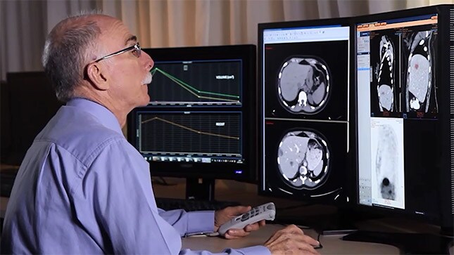
Dr. Raskin's experiences with MMTT.
We expect that as cancer becomes a chronic disease we will be doing multiple scans, maybe over the course of years, and they won’t all be CAT scans. There will be PET scans, there will be MRIs… We don’t know what is coming in the future, but we need one consistent way of tracking the tumors from time point to time point, regardless of the modality that we use to assess it.”
Stephen P. Raskin
M.D., Radiologist, Sheba Medical Center, Tel Ha-Shomer, Israel
How does it work
The Multi Modality Tumor Tracking application supports loading and comparison of previous and current patient datasets from CT, PET/CT, SPECT/CT, and MR.
Semi-automatic segmentation tools facilitate 2D and 3D segmentation of tumors and lymph nodes. Users can also use graphical tracking to chart the size of the tumor across different time points. After the measurements are completed, MMTT automatically performs the RECIST and WHO calculations. The MMTT application is explained in our recent white paper “Streamlined workflow for review and analysis of oncology patients”.
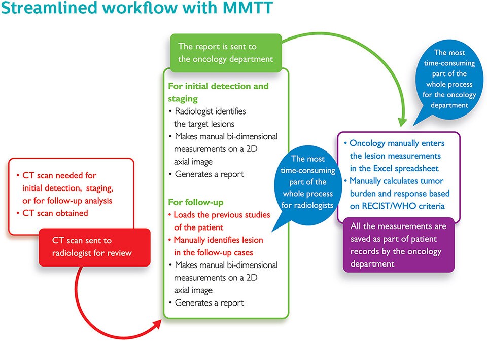
The above diagram shows how MMTT eliminates many time-consuming manual tasks in the oncology workflow.
Analyzing data
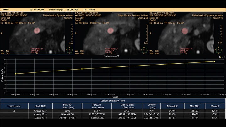
The MMTT application can also be used to analyze MR data. In the case shown on the right, a diffusion dataset is used to assess the size and activity of a lesion. The Apparent Diffusion Coefficient (ADC) values don’t change over the time period, whereas the size of the lesion appears to increase over the time frame.
