- Get step-by-step guidance to simplify CBCT acquisition
-
Get step-by-step guidance to simplify CBCT acquisition
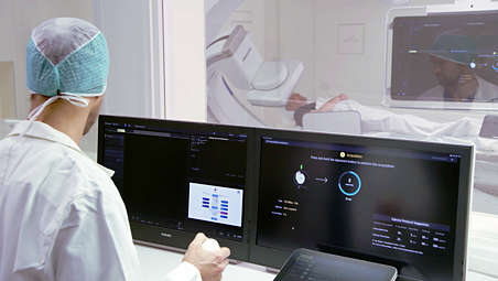
To help all clinical users achieve superb 3D images, regardless of their level of experience, SmartCT Soft Tissue provides step-by-step guidance and visual aids during acquisition. This includes room set-up, isocentering the system, and suggesting a suitable contrast injection and X-ray acquisition protocol[1]. - Interact with your CBCT image at tableside
-
Interact with your CBCT image at tableside
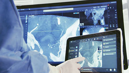
Once acquired, SmartCT Soft Tissue automatically displays the CBCT image on the touchscreen module and FlexVision within seconds for direct review at tableside[2]. - Control advanced 3D visualization and measurement tools at tableside
-
Control advanced 3D visualization and measurement tools at tableside
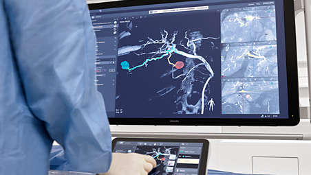
Using simple tablet gestures, you can carry out advanced measurements and visualizations on the touchscreen at tableside to study the disease in great detail. SmartCT 3D images can help reveal information not apparent on DSA images. This additional information may change diagnosis, treatment planning or treatment delivery, supporting better patient outcomes[3-5]. - CBCT Open for wider visualization
-
CBCT Open for wider visualization
SmartCT Soft Tissue offers the opportunity to acquire a CBCT using an open trajectory with start and stop positions of +55° to -185°, respectively. This protocol opens the arc to the left side of the patient, allowing for a wider translation of the angiographic table in this direction and shifting the isocenter of the C-arm to the right lateral side of the patient. This enables visualization of off-centered regions of interest (such as the periphery of the liver) in a single sweep[6]. The open arc trajectory can also ease imaging of larger patients. - CBCT Dual Phase CBCT for liver imaging
-
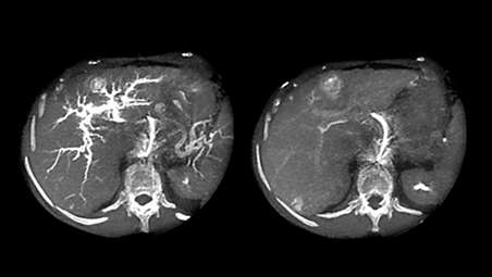
CBCT Dual Phase CBCT for liver imaging
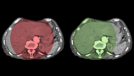
Dual Phase cone beam CT (CBCT) acquisition is an advanced capability of SmartCT Soft Tissue that allows two CBCT scans to be made automatically on the Azurion system with a user-defined interval and a single contrast injection. High-resolution, high-contrast images are reconstructed within seconds to support fast decisions during procedures. It is commonly used for TACE, where the first phase serves as an arterial phase and the second (delayed) phase shows contrast uptake in the lesions[7,8]. - CBCT Dual Phase for brain imaging
-
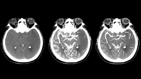
CBCT Dual Phase for brain imaging
Dual Phase CBCT is an increasingly used application for stroke diagnosis. A non-contract enhanced CBCT aids the visualization of soft tissue. - SmartCT Dual Viewer for easy reviewing and comparing of volumes
-
SmartCT Dual Viewer for easy reviewing and comparing of volumes
SmartCT Dual Viewer can take any two volumes from the Azurion System, display them side by side and overlay them to create fusion images to support the physician in assessment and diagnosis. The entire workflow from 3D acquisition protocol selection to image viewing, manipulation, image overlay and post-processing can be done in SmartCT. Dual Viewer in SmartCT allows the user to view and manipulate the volumes at tableside without breaking sterility using the touchscreen module (TSM) or mouse and keyboard.
Get step-by-step guidance to simplify CBCT acquisition
Get step-by-step guidance to simplify CBCT acquisition
Get step-by-step guidance to simplify CBCT acquisition
Interact with your CBCT image at tableside
Interact with your CBCT image at tableside
Interact with your CBCT image at tableside
Control advanced 3D visualization and measurement tools at tableside
Control advanced 3D visualization and measurement tools at tableside
Control advanced 3D visualization and measurement tools at tableside
CBCT Open for wider visualization
CBCT Open for wider visualization
CBCT Open for wider visualization
CBCT Dual Phase CBCT for liver imaging
CBCT Dual Phase CBCT for liver imaging
CBCT Dual Phase CBCT for liver imaging
CBCT Dual Phase for brain imaging
CBCT Dual Phase for brain imaging
CBCT Dual Phase for brain imaging
SmartCT Dual Viewer for easy reviewing and comparing of volumes

SmartCT Dual Viewer for easy reviewing and comparing of volumes
SmartCT Dual Viewer for easy reviewing and comparing of volumes
- Get step-by-step guidance to simplify CBCT acquisition
- Interact with your CBCT image at tableside
- Control advanced 3D visualization and measurement tools at tableside
- CBCT Open for wider visualization
- Get step-by-step guidance to simplify CBCT acquisition
-
Get step-by-step guidance to simplify CBCT acquisition
To help all clinical users achieve superb 3D images, regardless of their level of experience, SmartCT Soft Tissue provides step-by-step guidance and visual aids during acquisition. This includes room set-up, isocentering the system, and suggesting a suitable contrast injection and X-ray acquisition protocol[1]. - Interact with your CBCT image at tableside
-
Interact with your CBCT image at tableside
Once acquired, SmartCT Soft Tissue automatically displays the CBCT image on the touchscreen module and FlexVision within seconds for direct review at tableside[2]. - Control advanced 3D visualization and measurement tools at tableside
-
Control advanced 3D visualization and measurement tools at tableside
Using simple tablet gestures, you can carry out advanced measurements and visualizations on the touchscreen at tableside to study the disease in great detail. SmartCT 3D images can help reveal information not apparent on DSA images. This additional information may change diagnosis, treatment planning or treatment delivery, supporting better patient outcomes[3-5]. - CBCT Open for wider visualization
-
CBCT Open for wider visualization
SmartCT Soft Tissue offers the opportunity to acquire a CBCT using an open trajectory with start and stop positions of +55° to -185°, respectively. This protocol opens the arc to the left side of the patient, allowing for a wider translation of the angiographic table in this direction and shifting the isocenter of the C-arm to the right lateral side of the patient. This enables visualization of off-centered regions of interest (such as the periphery of the liver) in a single sweep[6]. The open arc trajectory can also ease imaging of larger patients. - CBCT Dual Phase CBCT for liver imaging
-
CBCT Dual Phase CBCT for liver imaging
Dual Phase cone beam CT (CBCT) acquisition is an advanced capability of SmartCT Soft Tissue that allows two CBCT scans to be made automatically on the Azurion system with a user-defined interval and a single contrast injection. High-resolution, high-contrast images are reconstructed within seconds to support fast decisions during procedures. It is commonly used for TACE, where the first phase serves as an arterial phase and the second (delayed) phase shows contrast uptake in the lesions[7,8]. - CBCT Dual Phase for brain imaging
-
CBCT Dual Phase for brain imaging
Dual Phase CBCT is an increasingly used application for stroke diagnosis. A non-contract enhanced CBCT aids the visualization of soft tissue. - SmartCT Dual Viewer for easy reviewing and comparing of volumes
-
SmartCT Dual Viewer for easy reviewing and comparing of volumes
SmartCT Dual Viewer can take any two volumes from the Azurion System, display them side by side and overlay them to create fusion images to support the physician in assessment and diagnosis. The entire workflow from 3D acquisition protocol selection to image viewing, manipulation, image overlay and post-processing can be done in SmartCT. Dual Viewer in SmartCT allows the user to view and manipulate the volumes at tableside without breaking sterility using the touchscreen module (TSM) or mouse and keyboard.
Get step-by-step guidance to simplify CBCT acquisition
Get step-by-step guidance to simplify CBCT acquisition
Get step-by-step guidance to simplify CBCT acquisition
Interact with your CBCT image at tableside
Interact with your CBCT image at tableside
Interact with your CBCT image at tableside
Control advanced 3D visualization and measurement tools at tableside
Control advanced 3D visualization and measurement tools at tableside
Control advanced 3D visualization and measurement tools at tableside
CBCT Open for wider visualization
CBCT Open for wider visualization
CBCT Open for wider visualization
CBCT Dual Phase CBCT for liver imaging
CBCT Dual Phase CBCT for liver imaging
CBCT Dual Phase CBCT for liver imaging
CBCT Dual Phase for brain imaging
CBCT Dual Phase for brain imaging
CBCT Dual Phase for brain imaging
SmartCT Dual Viewer for easy reviewing and comparing of volumes
SmartCT Dual Viewer for easy reviewing and comparing of volumes
SmartCT Dual Viewer for easy reviewing and comparing of volumes
Related products
Alternative products
-
SmartCT
- Simplifies 3D acquisition to empower clinical users to easily perform 3D imaging
- Enriches our outstanding 3D interventional tools with clear guidance
- 3D images are automatically displayed within seconds on the touch screen module
- Easily control and interact with advanced 3D visualization and measurement tools
View product
-
Azurion 7 B20/15
- Image Guided Therapy System Biplane with one 20" frontal and one 15" lateral flat detector
- Enhances certainty during neuro interventions like ischemic stroke and cerebral aneurysm treatment
- Experience a simple, smooth clinical workflow with the dedicated neuro features
View product
-
Azurion 7 M20
- Image Guided Therapy System Monoplane Ceiling/Floor Mounted with a 20" flat detector
- Covers a wide range of cardiac and vascular procedures to offer flexibility for multi-purpose use
- Control all relevant applications via the central touch screen module at tableside
- Parallel working enables you to get more done, leading to high throughput and fast exam turnover
View product
-
Azurion 5 M20
- Image Guided Therapy System Monoplane Ceiling/Floor Mounted with a 20” flat detector
- Covers a wide range of cardiac and vascular procedures to offer flexibility for multi-purpose use
- Control all relevant applications via the central touch screen module at tableside
- Parallel working enables you to get more done, leading to high throughput and fast exam turnover
View product
-
SmartCT Angio
- 3D visualization of bone and vessels from a single rotational angiographic X-ray acquisition
- Improve visibility of bone and vasculature in cerebral, abdominal and peripheral anatomies
- Acquire and interact with 3D imaging at table side
- 3D imaging can reveal information not apparent on DSA images
View product
-
SmartCT Roadmap
- Accurate and dynamic 3D guidance tool
- Real-time 3D view aids guidewire and catheter navigation through complex vessel structures
- Adapts to position changes in real-time
- Variable settings to enhance visualization
View product
-
SmartCT Vaso
- Step-by-step acquisition technique that can offer guidance to simplify 3D imaging
- Allows direct image inspection with advanced 3D visualization at table side
- Peri-procedure check of positioning of endovascular stents
View product
-
SmartCT Dual Viewer
- Simultaneously visualize any 2 volumes from Azurion of different sizes, at different procedure times
- View and manipulate datasets at tableside with the touchscreen module (TSM)
- Use the overlaid datasets as a 3D roadmap with fluoroscopy or to support with segmentation
View product
-
SmartCT Soft Tissue Helical
- Fast 10 secs helical trajectory acquisition to reduce motion artifacts
- Reconstruction software with cone beam, ring artifacts, bone beam, table scatter & vibration filters
- Automatic motion compensation functionality to salvage a CBCT with motion artifacts
- Metal artifact reduction algorithm to remove metal artifacts from the CBCT volume
- Workflow guidance with isocentering tool to aid 3D scan preparation
View product
-
SmartCT Dual Phase Cerebral
- Isocentering tool to aid with accurate positioning of the patient’s head
- Bolus Watch functionality to monitor contrast arrival and start CBCT acquisition at the right time
- Early phase contrast-enhanced cone beam CT (CBCT) to visualize large vessel occlusion
- Late-phase contrast-enhanced CBCT to visualize the presence of collateral vessels
- Dual Viewer to analyze the two CBCT volumes in an easy and intuitive way
View product


-
SmartCT
Philips Image Guided Therapy clinical application software SmartCT enriches our outstanding 3D interventional tools with clear guidance, designed to remove barriers to acquiring 3D images in the interventional lab. Once acquired, 3D images are automatically displayed within seconds on the touchscreen module in the corresponding rendering mode. On the same touchscreen, the user can easily control and interact with advanced 3D visualizations and measurement tools. SmartCT also brings advanced measurements and visualization to your fingertips for high image quality, supporting your diagnosis[1-3] and better patient treatment outcomes[4-6].
View product
-
Azurion 7 B20/15
Philips Azurion system with SmartCT allows you to perform a wide range of routine and complex interventional procedures easily and confidently with a unique user experience. Advanced capabilities integrated with an innovative system geometry support improved workflow, helping you to optimize your lab performance and provide superior care to your patients.
View product
-
Azurion 7 M20
Experience outstanding interventional cardiac and vascular performance on the Azurion 7 Series with 20'' flat detector. This industry-leading image-guided therapy solution enables your teams to benefit from superb consistency and efficiency as they perform diverse vascular and cardiac procedures. You can seamlessly control all relevant applications from a single touch screen a tableside, to help make fast, informed decisions in the sterile field.
View product
See all related products -
Azurion 5 M20
Experience outstanding interventional cardiac and vascular performance on the Azurion 7 Series with 20'' flat detector. This industry- leading image-guided therapy solution enables your teams to benefit from superb consistency and efficiency as they perform diverse vascular and cardiac procedures. You can seamlessly control all relevant applications from a single touch screen at tableside, to help make fast, informed decisions in the sterile field.
View product
-
SmartCT Angio
SmartCT Angio offers a 3D-RA (3D rotational angiography) acquisition protocol that provides extensive 3D visualization of bone and vessels based on a single contrast-enhanced rotational angiogram. Its high-resolution 3D reconstructions provide critical information about depth and the relationship of one vessel to another to support accurate assessment of bone and vasculature.
View product
-
SmartCT Roadmap
SmartCT Roadmap facilitates interventions by providing live 3D image guidance that can be segmented to emphasize target vessel and lesions, aiding guidewire and catheter navigation through complex vessel structures. All controlled via the touch screen at the table.
View product
-
SmartCT Vaso
SmartCT Vaso enables high-contrast and high-resolution imaging of cerebral vasculature based on a 3D rotational scan and an intra-arterial contrast injection. This technique enhances the visualization of endovascular stents, flow diverters, or other devices, as well as vessel morphology down to the perforator level.
View product
-
SmartCT Dual Viewer
SmartCT Dual Viewer can take any two volumes from the Azurion System, display them side by side and overlay them to create fusion images to support the physician in assessment and diagnosis and treatment planning (segmentation for example, manual feeder detection). Dual Viewer in SmartCT allows the user to view and manipulate the volumes at tableside without breaking sterility using the touchscreen module (TSM) or mouse and keyboard. There is also no need to switch applications because the entire workflow from 3D acquisition protocol selection to image viewing, manipulation, image overlay and postprocessing can be done in SmartCT and on the TSM.
View product
-
SmartCT Soft Tissue Helical
SmartCT Soft Tissue Helical creates CBCT images to help spot soft tissue changes in the Angio suite. The new protocol with dual-axis acquisition trajectory and improved reconstruction software results in images with improved image appearance compared to conventional cone beam acquisition techniques. SmartCT Soft Tissue Helical is our improved CBCT protocol for neurovascular care with a fast 8 secs trajectory, metal artifact and motion compensation algorithms to further improve image quality.
View product
-
SmartCT Dual Phase Cerebral
The Dual Phase Cerebral acquisition offers two consecutive contrast-enhanced cone beam CT scans of the brain. In the case of a stroke patient with a suspected large vessel occlusion, this type of acquisition allows identification of the vessel occlusion in the first phase, and the presence of collateral vessels in the second phase. This acquisition can be done with an intra-arterial as well as with intravenous contrast injection.
View product
- SmartCT R3.0 is subject to regulatory clearance and may not be available in all markets. Contact your sales representative for more details.
- 1. It is the operator’s responsibility to select the appropriate contrast agent depending on the clinical application. For more information about the indications for use of the contrast agent, refer to the instructions for use of the applicable contrast agent.
- 2. 3D reconstructions at higher resolution settings may take longer.
- 3. Ribo et al, Direct Transfer to Angiosuite to Reduce Door-To-Puncture Time in Thrombectomy for Acute Stroke, J Neurointerv Surg , 2018, 10 (3), 221-224
- 4. Fagan et al., MultiModality 3-dimensional image integration for Congenital Cardiac Catheterization. Methodist Debakey Cardiovasc J. 2014, 10 (2), 68-76
- 5. Hirotaka Hasegawa et al, Integration of rotational angiography enables better dose planning in Gamma Knife radiosurgery for brain arteriovenous malformations, J Neurosurg (Suppl) 129:17–25, 2018
- 6. Schernthaner et al., Feasibility of a Modified Cone-Beam CT Rotation Trajectory to Improve Liver Periphery Visualization during Transarterial Chemoembolization, Radiology, 2015carcinoma: comparison with intravenous contrast-enhanced, biphasic, dynamic MDCT. European Radiology. 22(4):872-9. DOI: 10.1007/s00330-011-2324-y
- 7. Higashihara, H., Osuga, K., Onishi, H., Nakamoto, A., Tsuboyama, T., Maeda, N., … Tomiyama, N. (2012). Diagnostic accuracy of C-arm CT during selective transcatheter angiography for hepatocellular carcinoma: comparison with intravenous contrast-enhanced, biphasic, dynamic MDCT. European Radiology. 22(4):872-9. DOI: 10.1007/s00330-011-2324-y.
- 8. Loffroy, R., Lin, M., Rao, P., Bhagat, N., Noordhoek, N., Radaelli, A., ... Geschwind, J.F. (2012). Comparing the detectability of hepatocellular carcinoma by C-arm dualphase cone-beam computed tomography during hepatic arteriography with conventional contrast-enhanced magnetic resonance imaging. CardioVascular and Interventional Radiology. 35(1):97-104. DOI: 10.1007/s00270-011-0118-x
- 9. Miyayama, S., Yamashiro, M., Hashimoto, M. et al. Comparison of Local Control in Transcatheter Arterial Chemoembolization of Hepatocellular Carcinoma ≤6 cm With or Without Intraprocedural Monitoring of the Embolized Area Using Cone-Beam Computed Tomography. Cardiovasc Intervent Radiol 37, 388–395