- 3D imaging at tableside
-
3D imaging at tableside
With the touchscreen, you can easily acquire 3D images and interact with SmartCT tools in a natural and effortless way. Once acquired, the SmartCT viewing application automatically opens your 3D image with the correct rendering and viewing tools on the touchscreen module. All tools work with touchscreen simplicity within the sterile area. - Reveal information not apparent on DSA images
-
Reveal information not apparent on DSA images
SmartCT offers advanced reconstruction techniques to improve anatomical visualization. Once acquired, you can manipulate advanced visualizations such as 3D MPR on the tableside touchscreen to evaluate the disease with great detail. SmartCT 3D images can help reveal information not apparent on DSA images, which can impact clinical decisions. - SmartCT Vessel Analysis supports treatment planning
-
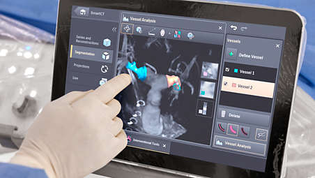
SmartCT Vessel Analysis supports treatment planning
Select two points of a vessel to quickly define a vessel path on a 3D volume. The path is automatically detected and rendered in different views to support easy inspection of vessel and device positioning with straightened, curved and cross-section reformats. The vessel dimensions are automatically extracted, and landing zones can be specified and later overlaid on live fluoroscopy. Finally, optimal projection angles for catheterization can be identified and later recalled with a single button press. - Easily perform two-point measurements onscreen
-
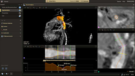
Easily perform two-point measurements onscreen
Quickly measure distance on a 2D or 3D image with the two-point measurement on the touchscreen. This can help you quickly check the trajectory to a target vessel, measure distances for stent deployment, measure the size of anatomy or identify a discrepancy to speed planning of the optimal treatment angle and aid navigation. - Quickly define any structure of interest
-
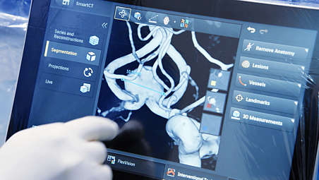
Quickly define any structure of interest
The SmartCT semi-automatic lesion segmentation tool allows you to easily define any structure of interest, measure its volume and remove anatomy to improve visualisation. - SmartCT Dual Viewer - 3D volume comparison and fusion solution
-
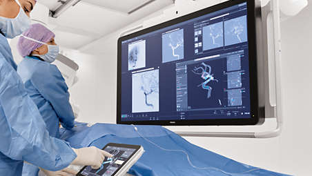
SmartCT Dual Viewer - 3D volume comparison and fusion solution
SmartCT Dual Viewer can take any two volumes from the Azurion System, display them side by side and overlay them to create fusion images to support the physician in assessment and diagnosis. The entire workflow from 3D acquisition protocol selection to image viewing, manipulation, image overlay and post-processing can be done in SmartCT. Dual Viewer in SmartCT allows the user to view and manipulate the volumes at tableside without breaking sterility using the touchscreen module (TSM) or mouse and keyboard.
3D imaging at tableside
3D imaging at tableside
3D imaging at tableside
Reveal information not apparent on DSA images
Reveal information not apparent on DSA images
Reveal information not apparent on DSA images
SmartCT Vessel Analysis supports treatment planning
SmartCT Vessel Analysis supports treatment planning
SmartCT Vessel Analysis supports treatment planning
Easily perform two-point measurements onscreen
Easily perform two-point measurements onscreen
Easily perform two-point measurements onscreen
Quickly define any structure of interest

Quickly define any structure of interest
Quickly define any structure of interest
SmartCT Dual Viewer - 3D volume comparison and fusion solution

SmartCT Dual Viewer - 3D volume comparison and fusion solution
SmartCT Dual Viewer - 3D volume comparison and fusion solution
- 3D imaging at tableside
- Reveal information not apparent on DSA images
- SmartCT Vessel Analysis supports treatment planning
- Easily perform two-point measurements onscreen
- 3D imaging at tableside
-
3D imaging at tableside
With the touchscreen, you can easily acquire 3D images and interact with SmartCT tools in a natural and effortless way. Once acquired, the SmartCT viewing application automatically opens your 3D image with the correct rendering and viewing tools on the touchscreen module. All tools work with touchscreen simplicity within the sterile area. - Reveal information not apparent on DSA images
-
Reveal information not apparent on DSA images
SmartCT offers advanced reconstruction techniques to improve anatomical visualization. Once acquired, you can manipulate advanced visualizations such as 3D MPR on the tableside touchscreen to evaluate the disease with great detail. SmartCT 3D images can help reveal information not apparent on DSA images, which can impact clinical decisions. - SmartCT Vessel Analysis supports treatment planning
-
SmartCT Vessel Analysis supports treatment planning
Select two points of a vessel to quickly define a vessel path on a 3D volume. The path is automatically detected and rendered in different views to support easy inspection of vessel and device positioning with straightened, curved and cross-section reformats. The vessel dimensions are automatically extracted, and landing zones can be specified and later overlaid on live fluoroscopy. Finally, optimal projection angles for catheterization can be identified and later recalled with a single button press. - Easily perform two-point measurements onscreen
-
Easily perform two-point measurements onscreen
Quickly measure distance on a 2D or 3D image with the two-point measurement on the touchscreen. This can help you quickly check the trajectory to a target vessel, measure distances for stent deployment, measure the size of anatomy or identify a discrepancy to speed planning of the optimal treatment angle and aid navigation. - Quickly define any structure of interest
-
Quickly define any structure of interest
The SmartCT semi-automatic lesion segmentation tool allows you to easily define any structure of interest, measure its volume and remove anatomy to improve visualisation. - SmartCT Dual Viewer - 3D volume comparison and fusion solution
-
SmartCT Dual Viewer - 3D volume comparison and fusion solution
SmartCT Dual Viewer can take any two volumes from the Azurion System, display them side by side and overlay them to create fusion images to support the physician in assessment and diagnosis. The entire workflow from 3D acquisition protocol selection to image viewing, manipulation, image overlay and post-processing can be done in SmartCT. Dual Viewer in SmartCT allows the user to view and manipulate the volumes at tableside without breaking sterility using the touchscreen module (TSM) or mouse and keyboard.
3D imaging at tableside
3D imaging at tableside
3D imaging at tableside
Reveal information not apparent on DSA images
Reveal information not apparent on DSA images
Reveal information not apparent on DSA images
SmartCT Vessel Analysis supports treatment planning
SmartCT Vessel Analysis supports treatment planning
SmartCT Vessel Analysis supports treatment planning
Easily perform two-point measurements onscreen
Easily perform two-point measurements onscreen
Easily perform two-point measurements onscreen
Quickly define any structure of interest
Quickly define any structure of interest
Quickly define any structure of interest
SmartCT Dual Viewer - 3D volume comparison and fusion solution
SmartCT Dual Viewer - 3D volume comparison and fusion solution
SmartCT Dual Viewer - 3D volume comparison and fusion solution
Related products
Alternative products
-
SmartCT
- Simplifies 3D acquisition to empower clinical users to easily perform 3D imaging
- Enriches our outstanding 3D interventional tools with clear guidance
- 3D images are automatically displayed within seconds on the touch screen module
- Easily control and interact with advanced 3D visualization and measurement tools
View product
-
Azurion 7 M12
- Image Guided Therapy System Monoplane Ceiling/Floor Mounted with a 12" flat detector
- Provides hi-res imaging over a large field of view, making it ideal for cardiac interventions
- Control all relevant applications via the central touch screen module at tableside
- Team members can work on multiple actions — at one or more workspots in the control and exam room
View product
-
Azurion 7 M20
- Image Guided Therapy System Monoplane Ceiling/Floor Mounted with a 20" flat detector
- Covers a wide range of cardiac and vascular procedures to offer flexibility for multi-purpose use
- Control all relevant applications via the central touch screen module at tableside
- Parallel working enables you to get more done, leading to high throughput and fast exam turnover
View product
-
Azurion 7 B12/12
- Image Guided Therapy System Biplane with two 12" flat detectors
- The C-arms can be independently positioned, for full patient access in anesthesiology/echocardiology
- Reveal critical anatomical information during congenital heart and electrophysiology procedures
View product
-
Azurion 7 B20/12
- Image Guided Therapy System Biplane with one 20" and one 12" flat detector
- Provides navigational precision for a wide range of challenging cardiac and vascular interventions
- Advanced interventional tools are seamlessly integrated to support your clinical workflow
View product
-
SmartCT Roadmap
- Accurate and dynamic 3D guidance tool
- Real-time 3D view aids guidewire and catheter navigation through complex vessel structures
- Adapts to position changes in real-time
- Variable settings to enhance visualization
View product
-
SmartCT Soft Tissue
- Step-by-step guidance technique to simplify cone beam computed tomography acquisition
- Interact with your CBCT image at table side on the touch screen module
- Access advanced 3D measurements at table side on the touch screen module
- Acquire a CBCT using open trajectory
View product
-
SmartCT Vaso
- Step-by-step acquisition technique that can offer guidance to simplify 3D imaging
- Allows direct image inspection with advanced 3D visualization at table side
- Peri-procedure check of positioning of endovascular stents
View product
-
SmartCT Dual Viewer
- Simultaneously visualize any 2 volumes from Azurion of different sizes, at different procedure times
- View and manipulate datasets at tableside with the touchscreen module (TSM)
- Use the overlaid datasets as a 3D roadmap with fluoroscopy or to support with segmentation
View product
-
VesselNavigator
- 3D image fusion technology for advanced endovascular procedures
- Supports navigation through complex vessel structures, enhancing clinical outcomes
- Reusing a pre-acquired CTA or MRA reduces the need for contrast enhanced runs
- Philips CTA Image Fusion Guidance may lead to shorter procedure times
View product


-
SmartCT
Philips Image Guided Therapy clinical application software SmartCT enriches our outstanding 3D interventional tools with clear guidance, designed to remove barriers to acquiring 3D images in the interventional lab. Once acquired, 3D images are automatically displayed within seconds on the touchscreen module in the corresponding rendering mode. On the same touchscreen, the user can easily control and interact with advanced 3D visualizations and measurement tools. SmartCT also brings advanced measurements and visualization to your fingertips for high image quality, supporting your diagnosis[1-3] and better patient treatment outcomes[4-6].
View product
-
Azurion 7 M12
Elevate your interventional cardiology capabilities with the Azurion 7 with 12'' flat detector. This high-performance image-guided therapy solution allows interventional teams to perform challenging cardiac interventions. Seamlessly control all relevant applications at tableside for a consistent user experience, excellent lab performance and patient care.
View product
-
Azurion 7 M20
Experience outstanding interventional cardiac and vascular performance on the Azurion 7 Series with 20'' flat detector. This industry-leading image-guided therapy solution enables your teams to benefit from superb consistency and efficiency as they perform diverse vascular and cardiac procedures. You can seamlessly control all relevant applications from a single touch screen a tableside, to help make fast, informed decisions in the sterile field.
View product
See all related products -
Azurion 7 B12/12
Discover amazing new possibilities for interventional cardiology, pediatric cardiology or electrophysiology with the Azurion 7 Series biplane with two 12'' detectors. This industry-leading Image Guided Therapy System allows you to easily and confidently perform procedures with consistent user experience, helping you optimize your lab performance and provide superior care. Seamlessly control all relevant applications from a single touch screen at tableside, to help make fast, informed decisions in the sterile field.
View product
-
Azurion 7 B20/12
Perform an array of cardiac and vascular interventions with singular precision and ease on the Azurion 7 biplane with one 20'' and one 12'' detector. This industry-leading image-guided therapy platform allows you to easily and confidently perform procedures with a unique user experience, helping you optimize your lab performance and provide superior care. Seamlessly control all relevant applications from a single touch screen at tableside, to help make fast, informed decisions in the sterile field.
View product
-
SmartCT Roadmap
SmartCT Roadmap facilitates interventions by providing live 3D image guidance that can be segmented to emphasize target vessel and lesions, aiding guidewire and catheter navigation through complex vessel structures. All controlled via the touch screen at the table.
View product
-
SmartCT Soft Tissue
SmartCT Soft Tissue offers a Cone Beam CT (CBCT) acquisition technique augmented with step-by-step guidance, Advanced 3D visualization and measurement tools all accessible on the touch screen module at table side. To support you in acquiring CBCT images first-time right and to streamline your workflow, you are guided through key steps. Once the CBCT scan is successfully performed, the acquired 3D image is automatically displayed in the SmartCT 3D visualization tool with the adequate rendering settings and the 3D measurement tools tailored for the selected 3D protocol.
View product
-
SmartCT Vaso
SmartCT Vaso enables high-contrast and high-resolution imaging of cerebral vasculature based on a 3D rotational scan and an intra-arterial contrast injection. This technique enhances the visualization of endovascular stents, flow diverters, or other devices, as well as vessel morphology down to the perforator level.
View product
-
SmartCT Dual Viewer
SmartCT Dual Viewer can take any two volumes from the Azurion System, display them side by side and overlay them to create fusion images to support the physician in assessment and diagnosis and treatment planning (segmentation for example, manual feeder detection). Dual Viewer in SmartCT allows the user to view and manipulate the volumes at tableside without breaking sterility using the touchscreen module (TSM) or mouse and keyboard. There is also no need to switch applications because the entire workflow from 3D acquisition protocol selection to image viewing, manipulation, image overlay and postprocessing can be done in SmartCT and on the TSM.
View product
-
VesselNavigator
VesselNavigator allows the reuse of 3D vascular anatomical information from existing CTA and MRA datasets as a 3D roadmap overlay on a live X-ray image. With its excellent visualization, VesselNavigator provides an intuitive, continuous 3D roadmap that guides you through the vasculature during the entire procedure.
View product
- SmartCT R3.0 is subject to regulatory clearance and may not be available in all markets. Contact your sales representative for more details.